Back Muscle Diagram / Upper Back Muscles Anatomy Anatomy Drawing Diagram. For example, some muscles located in the chest also help move the shoulders. The muscles of the back can be arranged into 3 categories based on their location: While muscles like the gluteals (in the thighs) are used any time we walk or climb a step, deep back muscles and abdominal muscles are usually not actively engaged during everyday activity. Superficial back muscles, intermediate back muscles and intrinsic back muscles.the intrinsic muscles are named as such because their embryological development begins in the back, oppose to the superficial and intermediate back muscles which develop elsewhere and are therefore classed as extrinsic muscles. The pelvis at the bottom of the back and the shoulders at the top of the back give the back.
Lower back muscle diagram anatomy does degenerative disc disease affect the lower back muscle? The back has a total of 40 muscles. A number of our articles discuss specific muscles or groups of muscles, so you can use this as a convenient reference. Back muscles chart, back muscles diagram and ligaments, back muscles diagram lats, back muscles diagram massage, upper back muscles chart, human muscles, back muscles. Start with 5 to 10 minutes of moderate cardio to get your blood pumping and start to awaken your muscles.
Back Muscle Anatomy Ankiweb from dl4.ankiweb.net Take a look at the following muscle back diagrams that we posted below to see more details of the muscle anatomy diagram. Another common cause of lower back and hip pain is disc injury. Superficial, intermediate, deep and deepest layers.these muscles lie on each side of the vertebral column, deep to the thoracolumbar fascia they span the entire length of the vertebral column, extending from the cranium to the pelvis Related posts of muscles of the lower back and buttocks diagram muscle anatomy multiple choice. The pelvis at the bottom of the back and the shoulders at the top of the back give the back. Some of the links in the post above are affiliate links.. Nerves in your lower back. Click at least one area of the body marked by a red arrow.
Anatomynote.com found anatomy of back muscles diagram from plenty of anatomical pictures on the internet.
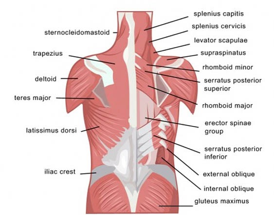
Introduction to musculoskeletal pathologies of the low back and pelvis. The deltoid, teres major, teres minor, infraspinatus, supraspinatus (not shown) and subscapularis muscles (not shown) all extend from the scapula to the humerus and act on the shoulder joint. The deep back muscles, also called intrinsic or true back muscles, consist of four layers of muscles: Deep back muscles diagram the superficial layer contains the splenius cervicis and splenius capitis muscles. Your back pain map can be printed after selecting symptoms from at least one area of the body. The muscles of the back can be arranged into 3 categories based on their location: Below you'll see diagrams along with the names of the back muscles that may be the cause of your pain. Most of the time, back muscle pain is diagnosed then treated with little more than a prescription of rest, painkillers and muscle relaxants. The trapezius is a broad, flat and triangular muscle. Click at least one area of the body marked by a red arrow. The muscles of the lower back help stabilize, rotate, flex, and extend the spinal column, which is a bony tower of 24 vertebrae that gives the body structure and houses the spinal cord. For more anatomy content please follow us and visit our website: The intermediate layer contains the erector spinae muscles, whose many functions include the extension and lateral flexion of the spine, head and neck.
Introduction to musculoskeletal pathologies of the low back and pelvis. The back consists of the spine, spinal cord, muscles, ligaments, and nerves. Click at least one area of the body marked by a red arrow. That can be a big step in gaining better control over your pain and improving your quality of life. Select all symptoms that apply in the menu that appears.
Back Strains And Sprains Back Pain Treatment from www.backpaintreatment.com Back muscles chart, back muscles diagram and ligaments, back muscles diagram lats, back muscles diagram massage, upper back muscles chart, human muscles, back muscles. The trapezius is a broad, flat and triangular muscle. The part of the nerve that emerges out of the spine is called the nerve root. Likewise, there are muscles in other parts of the body that help support and move the spine. Browse 195 back muscles diagram stock photos and images available, or start a new search to explore more stock photos and images. Below you'll see diagrams along with the names of the back muscles that may be the cause of your pain. The deep muscles develop embryologically in the back, and are thus described as intrinsic muscles. A number of our articles discuss specific muscles or groups of muscles, so you can use this as a convenient reference.
These structures work together to support the body, enable a range of movements, and send messages from the.
Lower back muscle diagram anatomy does degenerative disc disease affect the lower back muscle? Related posts of muscles of the lower back and buttocks diagram muscle anatomy multiple choice. That can be a big step in gaining better control over your pain and improving your quality of life. The pelvis at the bottom of the back and the shoulders at the top of the back give the back. Muscle diagram of the back (posterior) & front (anterior) this labeled human muscular system chart illustrates the major muscle groups in the back (posterior) view and the front (anterior) view. How many muscles are in the back? Superficial, intermediate, deep and deepest layers.these muscles lie on each side of the vertebral column, deep to the thoracolumbar fascia they span the entire length of the vertebral column, extending from the cranium to the pelvis Nerves in your lower back. The deltoid, teres major, teres minor, infraspinatus, supraspinatus (not shown) and subscapularis muscles (not shown) all extend from the scapula to the humerus and act on the shoulder joint. Start with 5 to 10 minutes of moderate cardio to get your blood pumping and start to awaken your muscles. The superficial and intermediate muscles do not develop in the back, and are classified as extrinsic muscles. Muscle diagrams are a great way to get an overview of all of the muscles within a body region. The deep back muscles, also called intrinsic or true back muscles, consist of four layers of muscles:
Daniel nelson on january 1, 2019 2 comments 🔥! Creatine research more than a sports supplement read more…. The deep muscles develop embryologically in the back, and are thus described as intrinsic muscles. The fibres attach to the clavicle, acromion and the scapula spine. Others, like sumo deadlifts, have been shown in emg studies—and in the trenches—to focus more on other muscle groups than the back.
Upper Back Muscles Medical Art Library from medicalartlibrary.com Creatine is now proving to be one of the most potent muscle growth accelerators giving excellent muscle mass increase and phenomenal strength increases order yours today. Superficial, intermediate, deep and deepest layers.these muscles lie on each side of the vertebral column, deep to the thoracolumbar fascia they span the entire length of the vertebral column, extending from the cranium to the pelvis A number of our articles discuss specific muscles or groups of muscles, so you can use this as a convenient reference. The superficial and intermediate muscles do not develop in the back, and are classified as extrinsic muscles. The human back extends from the buttocks to the posterior portion of the neck and shoulders. Your back pain map can be printed after selecting symptoms from at least one area of the body. The back has a total of 40 muscles. Anatomynote.com found anatomy of back muscles diagram from plenty of anatomical pictures on the internet.
Anatomynote.com found anatomy of back muscles diagram from plenty of anatomical pictures on the internet.
See back muscles and low back pain. Others, like sumo deadlifts, have been shown in emg studies—and in the trenches—to focus more on other muscle groups than the back. (the short head of the biceps femoris even. The deep muscles develop embryologically in the back, and are thus described as intrinsic muscles. The trapezius is a broad, flat and triangular muscle. The superficial and intermediate muscles do not develop in the back, and are classified as extrinsic muscles. Start with 5 to 10 minutes of moderate cardio to get your blood pumping and start to awaken your muscles. The fibres attach to the clavicle, acromion and the scapula spine. Related posts of muscles of the lower back and buttocks diagram muscle anatomy multiple choice. The deep back muscles, also called intrinsic or true back muscles, consist of four layers of muscles: Creatine research more than a sports supplement read more…. Your back pain map can be printed after selecting symptoms from at least one area of the body. These structures work together to support the body, enable a range of movements, and send messages from the.